LES LUNDIS DE SAINT-ANTOINE
Bâtiment Kourilsky - 11h–12h
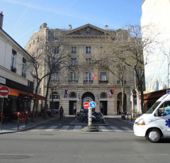
Salle des Conférences (Rez de Chaussée),
184 rue du Faubourg Saint-Antoine, Paris
***
LUNDI 23 NOVEMBRE 2026
Where you meet the microbiota: Peyer's patch LysoDCs instruct protective commensal-specific T cell programs
Oliveira Corrêa, RENAN
Postdoctoral Researcher, Institut IMAGINE, (Dr. Bana Jabri / Dr. Nadine Cerf-Bensussan)
invited by Pistorio Valeria Jérémie GAUTHERON's team François Delhommeau (Team Jérémie GAUTHERON
(valeria.pistorio@inserm.fr)
***
Segmented filamentous bacteria (SFB) potently induce intestinal Th17 immunity, yet how distinct gut-associated lymphoid tissues shape commensal-specific CD4 T-cell programs remains unclear. Using gnotobiotic models, SFB-specific TCR transgenic cells, and single-cell transcriptomic and TCR repertoire analyses, we show that Peyer's patches (PPs) and mesenteric lymph nodes (MLNs) act as distinct instructive niches during early SFB colonization. We identify monocyte-derived LysoDCs in PPs as specialized antigen-presenting cells that capture luminal SFB in the subepithelial dome, migrate to interfollicular regions, and engage SFB-specific CD4 T cells, being uniquely able to prime them from endogenously acquired antigen. In mice lacking CCR2-dependent LysoDCs, SFB-specific T cells accumulated and activated normally in PPs but showed impaired Th17 commitment and reduced type 1 regulatory (Tr1)-like programming in the small intestine. PP priming induced activated, clonally expanded Th17-biased populations with tissue-retention programs, glycolytic rewiring and Tr1-signature gene expression. In contrast, MLN responses were CCR2-independent and generated recirculating, less differentiated, predominantly singleton clonotypes. Functionally, PP-primed cells better preserved barrier integrity and limited pathology following transfer into Citrobacter rodentium-infected lymphopenic hosts, whereas MLN-primed cells did not. These findings establish the anatomical site of commensal priming as a major determinant of CD4 T-cell fate and protective function.
INSERM
Kourilsky Building
34 rue Crozatier - 75012 Paris
France
Sorbonne Université Medicine
Saint-Antoine Site
27 rue Chaligny - 75012 Paris
France